
Medical review status: This article is based on established medical references but has not been medically reviewed by a named healthcare professional.
Medical disclaimer: An ear lump cannot be diagnosed reliably from an online description alone. Seek professional evaluation for a new, persistent, growing, bleeding, crusting, infected, or otherwise changing lesion.
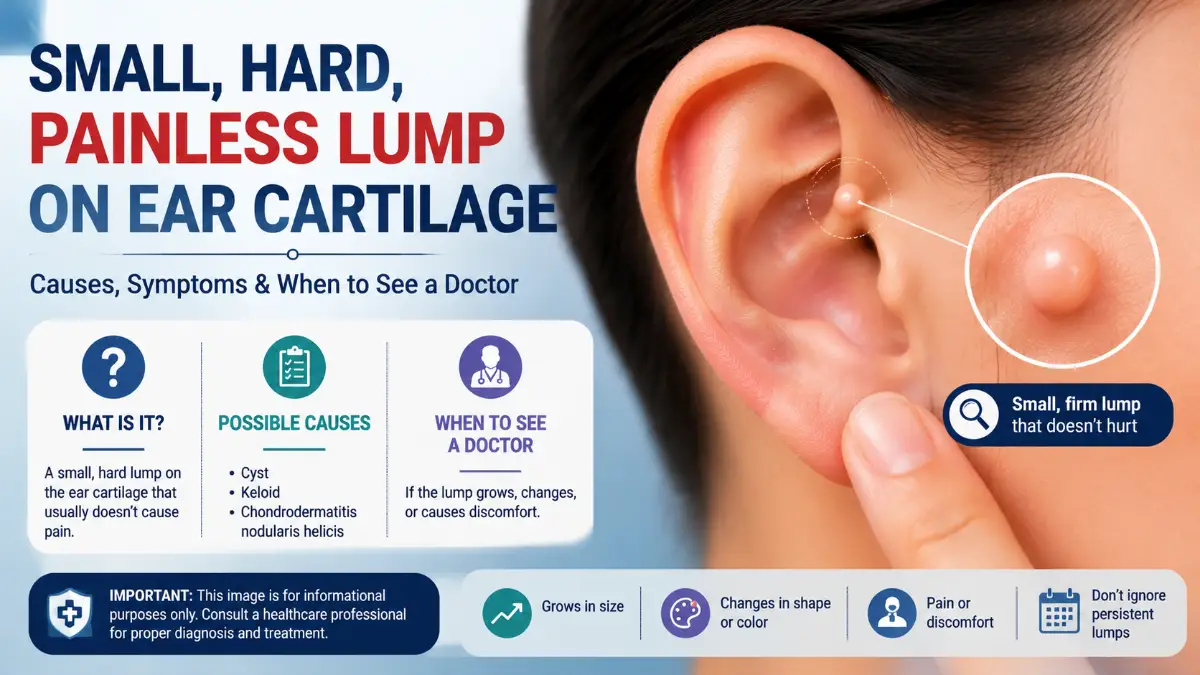
Finding a small hard painless lump on ear cartilage can be confusing because several very different things may feel similar when you touch them.
Some lumps arise from the skin covering the ear. Others are scars, fluid collections within cartilage, normal anatomical prominences, or less commonly skin growths that require investigation. The absence of pain can be useful information, but it does not prove that the lump is harmless.
A practical way to narrow the possibilities is to answer three questions:
- Exactly where on the ear is the lump?
- Does it move with the skin or feel fixed to the cartilage?
- What happened before you noticed it-piercing, pressure, trauma, or nothing obvious?
Those clues can help you describe the problem accurately to a doctor, but they should not be used to make a definite diagnosis at home.
First, Make Sure the Lump Is Actually on Ear Cartilage
The visible outer ear is called the auricle or pinna.
Most of it is supported by elastic cartilage, including the:
- helix,
- antihelix,
- tragus,
- antitragus,
- concha.
The earlobe does not contain cartilage.
That distinction is important because a lump within the soft earlobe may have different likely causes from one positioned over the upper helix or antihelix. The outer ear also has naturally firm ridges and projections. Some normal anatomical variations can feel surprisingly hard, particularly when a person notices them for the first time.
A quick location guide
Also Read: Knee Cartilage: Damage, Repair, Exercises & Treatment Guide
| Location | What to consider |
| Outer rim or helix | Normal cartilage, skin lesion, CNH, cyst |
| Inner ridge or antihelix | CNH, cartilage swelling, pseudocyst |
| Upper flat cartilage | Auricular pseudocyst, trauma-related change |
| Old piercing site | Keloid or other scar tissue |
| Earlobe | Cyst, scar or piercing-related lump rather than cartilage |
| Behind the ear | Different group of causes, not technically an ear-cartilage lump |
Professional external-link hint: Link the phrase “external ear anatomy” to the NCBI/StatPearls external-ear anatomy reference.
What Can Cause a Small Hard Painless Lump on Ear Cartilage?
No single condition accounts for every painless ear bump. The most useful possibilities to understand are normal cartilage anatomy, epidermoid cysts, piercing scars or keloids, auricular pseudocysts, and certain skin lesions.
1. A Normal Cartilage Prominence
One overlooked possibility is that the “lump” is simply part of the ear’s natural structure.
Human ears vary considerably in:
- cartilage thickness,
- ridge prominence,
- symmetry,
- helix shape,
- antihelix shape.
A known anatomical variant called Darwin’s tubercle can produce a small firm prominence on the helix.
A normal cartilage feature becomes more plausible if it:
- appears to have existed for a long time,
- feels exactly as hard as the surrounding cartilage,
- has completely normal skin over it,
- does not enlarge,
- does not bleed or crust,
- causes no tenderness.
Compare the area gently with the same position on the opposite ear, but remember that normal ears are not always perfectly symmetrical. A newly appearing or changing bump should not automatically be dismissed as normal anatomy.
2. An Epidermoid Cyst Under the Ear Skin
An epidermoid cyst is a benign sac beneath the skin containing keratin. Although people sometimes call these “sebaceous cysts,” epidermoid cysts do not actually arise from sebaceous glands.
They may appear as:
- a smooth round bump,
- a skin-colored nodule,
- a movable lump just under the skin,
- a lesion with a tiny central opening or punctum.
An uncomplicated cyst can remain painless for a long time.
If it becomes inflamed or ruptures, it may instead become:
- red,
- swollen,
- tender,
- warm,
- larger.
How a cyst may differ from cartilage
One useful clue is mobility. A superficial cyst may move slightly beneath the skin, while a true cartilage prominence usually feels more integrated into the structure of the ear. That distinction is not reliable enough for self-diagnosis, however. Professional external-link hint: Link “epidermoid cyst” to the NCBI/StatPearls epidermal inclusion cyst reference.
Read More: Human Torso Anatomy: Complete Guide to Structure, Organs, Bones & Muscles
3. A Keloid or Raised Scar From a Piercing
If you have ever had the area pierced, scar tissue should move much higher on the list of possibilities. A keloid is a type of raised scar that grows beyond the original boundaries of the skin injury.
Ear piercing is a well-established trigger.
A keloid may:
- feel firm or rubbery,
- develop gradually,
- continue growing after the original wound has healed,
- extend beyond the piercing hole,
- be painless once mature,
- sometimes itch, burn, or become tender while growing.
Keloids are particularly important to distinguish from ordinary “piercing bumps.”
Not every raised piercing scar is a keloid.
A useful history question
Ask yourself:
Is the center of the lump exactly where the piercing needle originally passed through the ear? If yes, a scar-related process becomes more plausible. Do not attempt to cut or tie off an ear keloid yourself. Dermatologists have several treatment approaches, and keloids can recur after treatment. Professional external-link hint: Link “keloid after ear piercing” to the American Academy of Dermatology‘s keloid guidance.
4. Auricular Pseudocyst
An auricular pseudocyst is different from a skin cyst. It involves a fluid-containing space forming within the cartilage of the external ear. DermNet describes auricular pseudocysts as rare and usually asymptomatic. They may arise spontaneously or after trauma.
They commonly affect the upper part of the ear.
The swelling may appear:
- smooth,
- rounded,
- painless,
- non-red,
- contained within the cartilage.
Because there is fluid involved, some pseudocysts feel less rock-hard than a solid cartilage projection.
Why this distinction matters
An epidermoid cyst forms in the skin or tissue underneath it. An auricular pseudocyst forms within the cartilage itself. Treatment may therefore be quite different. Repeatedly draining a suspected pseudocyst yourself is not recommended. Improper treatment can increase the risk of infection, recurrence, or ear deformity. Professional external-link hint: Link “auricular pseudocyst” to DermNet’s auricular pseudocyst reference.
5. Could It Be Chondrodermatitis Nodularis Helicis?
Chondrodermatitis nodularis helicis, often abbreviated CNH, commonly appears on the helix or antihelix.
It can produce a small firm nodule with:
- crusting,
- scaling,
- central ulceration,
- redness.
However, there is an important mismatch with the keyword being discussed here:
CNH is usually painful or tender.
People may especially notice pain when:
- sleeping on the affected side,
- pressing the ear,
- wearing headphones,
- exposing the ear to cold.
Therefore, a completely painless bump is less typical of classic CNH. That does not mean symptoms alone can rule it out. CNH can sometimes resemble other ear lesions, which is why persistent or unusual lesions may require a dermatologist’s assessment. Professional external-link hint: Link “chondrodermatitis nodularis helicis” to DermNet.
Does “Hard” Tell You What the Lump Is?
Not necessarily. People use the word hard to describe several different textures.
A useful distinction is:
Bone-hard or cartilage-hard
Feels as though it is part of the ear’s framework.
This may suggest:
- a normal cartilage projection,
- longstanding cartilage alteration,
- another structure involving the cartilage.
Firm but movable
Feels solid but shifts somewhat underneath the skin.
This may fit:
- an epidermoid cyst,
- another superficial skin nodule.
Firm scar-like tissue
Often occurs precisely at an old piercing or wound.
This may fit:
- a keloid,
- hypertrophic scar.
Rounded swelling with some give
A lesion that feels less solid may raise consideration of:
- a cyst,
- pseudocyst,
- another fluid-containing process.
Touch alone cannot reliably identify the cause.
Does “Painless” Mean It Cannot Be Cancer?
No.Pain is not a reliable way to distinguish benign from malignant skin lesions. Most small painless ear bumps are not automatically cancer, but skin cancers can sometimes be painless. The outer ear is also a sun-exposed area.
Squamous cell carcinoma can sometimes appear as a:
- firm growth,
- rough or scaly patch,
- crusted lesion,
- enlarging bump,
- sore that does not heal.
A changing lesion on the ear therefore deserves more attention than a tiny stable bump that has remained identical for years.
Features that deserve medical assessment
Arrange an examination if the lump:
- keeps enlarging,
- repeatedly crusts,
- bleeds without being picked,
- develops an ulcer,
- becomes persistently scaly,
- changes color,
- develops irregular edges,
- creates a non-healing sore,
- begins distorting the shape of the ear.
Professional external-link hint: Link “squamous cell carcinoma signs” to the American Academy of Dermatology rather than a commercial health blog.
Read More: Emergency Dentist: A Fast Action Plan for Tooth Pain, Swelling and Dental Injuries
Use This 5-Clue Method Before Your Appointment
Instead of repeatedly touching the bump, record these five characteristics.
1. Location
Write down whether it is on the:
- helix,
- antihelix,
- concha,
- tragus,
- piercing site,
- earlobe,
- skin behind the ear.
A photo of labeled ear anatomy can help you identify the correct term.
2. Size
Measure the approximate width with a ruler.
Avoid repeatedly estimating it from memory.
3. Surface
Look for:
- completely normal skin,
- scaling,
- crust,
- tiny central opening,
- ulceration,
- discoloration.
4. Movement
Without squeezing hard, notice whether the bump appears to move separately from the ear cartilage.
5. Change over time
Take a clear photograph under similar lighting. Recording the date can make it easier to determine whether it is actually changing.
Questions That Can Narrow the Possibilities
A clinician may want to know:
- How long has the lump been there?
- Did it appear suddenly or gradually?
- Has it become larger?
- Was the ear previously pierced?
- Have you injured that part of the ear?
- Do you usually sleep on that side?
- Do headphones, helmets, or hearing aids press on it?
- Has it ever drained fluid?
- Has it ever bled?
- Does it become tender when pressed?
- Do you have similar lumps elsewhere?
- Have you had substantial lifetime sun exposure?
The answers may be more informative than simply saying that the lump feels “hard.”
Hard Ear Lump After a Cartilage Piercing
A piercing changes the differential diagnosis significantly.
A bump following a piercing can represent:
- temporary irritation,
- excessive scar formation,
- a keloid,
- infection,
- another localized reaction.
Signs that sugg
est infection instead of a quiet scar
Watch for:
- increasing pain,
- spreading redness,
- warmth,
- marked swelling,
- yellow or green drainage,
- fever,
- feeling unwell.
Cartilage infections deserve particular attention because the ear’s cartilage depends on surrounding tissue for its blood supply and serious infection can damage the ear. Seek medical care rather than attempting to drain an infected cartilage piercing yourself.
Should You Pop or Pierce the Lump?
No. A small hard painless lump on ear cartilage is not necessarily a pimple.
Puncturing an unexplained lesion can:
- introduce bacteria,
- trigger inflammation,
- create bleeding,
- damage cartilage,
- cause scarring,
- make the lesion harder to assess.
Do not use:
- sewing needles,
- pins,
- home lancing tools,
- wart acid,
- harsh chemical peels,
- repeated squeezing.
Knowing the diagnosis should come before trying to remove the lump.
When Is an Ear Lump More Reassuring?
Certain characteristics may be relatively reassuring, although they never provide a guarantee.
Examples include a bump that:
- has been unchanged for years,
- remains very small,
- has smooth normal skin,
- has no bleeding,
- has no ulceration,
- has no discharge,
- causes no symptoms,
- corresponds closely to a normal cartilage structure.
Even then, an in-person examination is reasonable if you are uncertain whether the lump is new.
When Should You Make a Routine Doctor’s Appointment?
Consider arranging a non-emergency appointment if the lump is:
- new and does not disappear,
- gradually enlarging,
- of uncertain cause,
- cosmetically concerning,
- located at an old piercing and continues growing,
- repeatedly becoming inflamed,
- different from the same area on the opposite ear.
A persistent unexplained skin lesion is better assessed than repeatedly searched online.
When Should It Be Checked More Promptly?
Seek faster medical advice for:
- rapid enlargement,
- spontaneous bleeding,
- ulceration,
- significant redness or warmth,
- pus,
- worsening pain,
- fever,
- spreading swelling,
- significant ear deformity.
A suspicious or changing skin growth also deserves timely dermatological assessment.
Dermatologist, ENT, or Primary-Care Doctor?
The best starting point depends on what the lesion appears to involve.
| Situation | Useful clinician |
| Uncertain small lump | Primary-care clinician |
| Surface skin growth, crust or pigment change | Dermatologist |
| Possible cyst | Dermatologist or primary care |
| Suspected keloid | Dermatologist |
| Possible cartilage pseudocyst | ENT or dermatologist |
| Ear canal involvement | ENT |
| Possible skin cancer | Dermatologist |
| Significant cartilage infection | Prompt medical/ENT assessment |
A clinician may refer you if the diagnosis falls outside their specialty.
How Is a Persistent Ear Lump Diagnosed?
Many outer-ear lumps can initially be assessed through:
- medical history,
- visual examination,
- palpation.
A dermatologist may use dermoscopy, which provides a magnified view of structures within the skin.
A biopsy may be considered if:
- the diagnosis is uncertain,
- a lesion looks suspicious,
- a persistent nodule resembles more than one condition.
Not every small ear bump requires scans or imaging. Testing should be selected according to what a clinician finds during examination.
Treatment Depends on the Diagnosis
There is no universal treatment for a painless cartilage lump.
| Possible cause | General management approach |
| Normal anatomical prominence | Usually no treatment |
| Stable epidermoid cyst | Observation or elective removal when appropriate |
| Keloid | Dermatologist-directed scar treatment |
| Auricular pseudocyst | Clinician-directed drainage/compression or another procedure |
| CNH | Pressure reduction and medical/procedural treatment |
| Infection | Medical treatment based on severity and cause |
| Suspicious skin growth | Examination and biopsy/removal when indicated |
This is why trying a random treatment before diagnosis is often counterproductive.
Frequently Asked Questions
What is the most common cause of a small hard painless lump on ear cartilage?
There is no single cause that can be identified from those three characteristics alone. Normal cartilage anatomy, cysts and scar tissue are among the possibilities. Location, mobility, piercing history and surface appearance provide important additional clues.
Why does my ear have a hard bump but the other ear does not?
Normal ears are not perfectly symmetrical. A cartilage ridge or anatomical variation can be more prominent on one side. A truly new asymmetric lump, however, should be monitored and assessed if persistent.
Can a cyst on the ear be painless?
Yes. Epidermoid cysts are often asymptomatic until they become inflamed or rupture.
Can a keloid be painless?
Yes. Keloids can cause itching, burning or tenderness while growing, but some mature keloids cause little or no discomfort.
What if the hard lump is exactly where my cartilage piercing was?
Piercing-related scar tissue or a keloid becomes more plausible. Redness, warmth, worsening pain or drainage instead raises concern about infection.
Is a painless ear bump likely to be CNH?
Classic CNH is usually painful or tender, especially with pressure. A completely painless lump is therefore less characteristic, although only an examination can determine the diagnosis.
Can skin cancer occur on the ear without pain?
Yes. Lack of pain does not exclude skin cancer. Growth, bleeding, crusting, ulceration or a sore that does not heal are more useful reasons to seek evaluation.
Can I watch a small ear bump for changes?
Photographing and measuring a stable-looking bump can help document changes, but a new unexplained lesion should not be monitored indefinitely if it persists or changes.
Should I squeeze the lump to see if anything comes out?
No. Squeezing or puncturing an undiagnosed ear lump can cause inflammation, infection and scarring.
